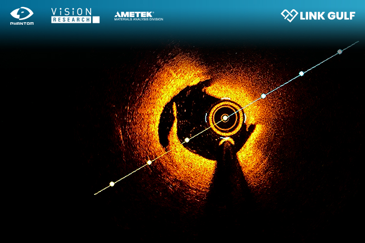
Optical coherence tomography (OCT) is a non-invasive and non-destructive imaging technique that provides a two or three-dimensional in vivo visualisation of tissue structures. Filling a gap between ultrasound’s imaging depth and confocal microscopy’s spatial resolution, OCT has revealed insights in biomedical applications like ophthalmology and laryngology.
Recently, researchers have begun integrating high-speed cameras into OCT instruments and systems. Taking advantage of the high throughput of Phantom cameras, these new imaging systems deliver exceptional data acquisition in milliseconds, surpassing the speed of traditional OCT scanning methods by a wide margin.
The newest class of OCT technology is Fourier Domain OCT (FD-OCT). This uses low-coherence interferometry to compare the optical path length delays between a sample and a reference light source. There are two types of FD-OCT systems — spectral domain (SD-OCT) and swept source (SS-OCT).
These systems differ in their light sources and detection methods, but both rely on interferometry. Interferometry uses the light scattered back due to refractive index variations within a sample. It combines light that has travelled with a fixed reference length. An interferometer detects the combined light and the resulting interferogram’s frequency represents the depths of the reflectors in the sample. Applying the Fourier transform to the interferogram provides a depth reflectivity profile of the sample at that point.
To obtain a three-dimensional volume of the sample, the system scans the sample in one direction to generate a two-dimensional cross-section. Scanning in a perpendicular direction, additional cross-sections are obtained and then combined to form a three-dimensional dataset.
While conventional approaches rely on single-point detectors and galvanometer scanners, the new integrated system eliminates the need for mechanical scanning. It also streamlines the imaging process. An additional advantage, combining high-speed OCT-based systems with newer Phantom Machine Vision cameras, provides ultra-long record times and real-time image analysis.
Data acquisition is enhanced to unprecedented speeds by incorporating holographic OCT, which features a swept-source laser on a sensor array. In application, researchers have unveiled an advanced, real-time holographic retinal blood flow imaging instrument. All powered by a Phantom S710 streaming camera. The system enables real-time Doppler image calculations from a continuous stream of high-resolution interferograms. All while recording at 4,000 frames per second.
Using Fresnel transformation and local principal component analysis filtering, the image rendering process achieves a temporal resolution of 125 fps. These results demonstrate successful real-time imaging of blood flow in iris arteries and veins. As a result of the system’s versatility, researchers can identify crucial anatomical landmarks. Such as observing arterial pulse waves and venous flow in vessels of varying sizes.
Given the ability to integrate Phantom high-speed cameras into interferometry workflows. This means that new, noninvasive medical imaging techniques are possible.
Learn more about our machine vision cameras.
Discover more here: View the Phantom Machine Vision Product Range >>
Phantom Cameras Website: phantomhighspeed.com
